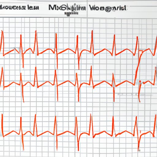
Exploring the Basics of an Echo Cardio Gram
An echo cardiogram (also known as an echocardiogram or ECG) is a type of imaging test used to examine the structure and function of the heart. It uses sound waves to create detailed images of the heart and its valves. An echo cardiogram is an important diagnostic tool for detecting and monitoring various types of heart conditions, such as congenital heart defects, coronary artery disease, and heart valve problems.
How it Works
During an echo cardiogram, an ultrasound transducer is placed on the chest wall. This device emits sound waves that are reflected back by the heart and other tissues in the chest. The echoes are captured by the transducer and sent to a computer, which converts them into a real-time image of the heart. This image can be viewed on a monitor and used to assess the size and shape of the heart, as well as the motion of its walls and valves.

How an Echo Cardio Gram Helps Doctors Diagnose Heart Conditions
The information gathered from an echo cardiogram helps doctors diagnose a variety of heart conditions. For example, it can detect problems with the heart’s chambers and valves, as well as determine the thickness of the heart walls. It can also show areas of poor blood flow due to blockages, and detect the presence of fluid in the lungs. In addition, an echo cardiogram can measure the pumping strength of the heart, and provide information about the size and shape of the major arteries that supply the heart.
Types of Tests Used
Doctors typically use several different types of tests to diagnose heart conditions. These include electrocardiograms (ECGs), chest X-rays, and cardiac catheterizations. However, an echo cardiogram is often the most accurate and reliable test for diagnosing many types of heart problems. It provides detailed images of the heart that can be used to accurately diagnose and monitor various heart conditions.
Benefits to Patients
An echo cardiogram can provide important information to patients about their heart health. It can help them understand what treatments might be necessary, and even provide peace of mind if no serious problems are found. Additionally, it can be used to monitor the progress of certain heart conditions over time.
What to Expect During an Echo Cardio Gram Procedure
An echo cardiogram is generally a safe and painless procedure. However, there may be some discomfort associated with the placement of the transducer on the chest wall. Here is what to expect during an echo cardiogram procedure:
Preparing for the Test
Before the test, you should remove any jewelry or clothing that could interfere with the transducer. You may also be asked to lie down on a bed or table, and the technician may place electrodes on your chest to monitor your heart rhythm. Additionally, you may need to take a deep breath and hold it for a few seconds while the technician takes pictures of your heart.
The Procedure
During the procedure, the technician will use the ultrasound transducer to transmit sound waves through your chest wall. These sound waves will be reflected by the heart and other tissues in your chest, creating an image of your heart that can be seen on a monitor. The entire procedure typically takes about 30 minutes.
After the Procedure
After the procedure, you may experience some minor discomfort in your chest. However, this should resolve quickly. Your doctor or technician will discuss the results of the test with you, and may recommend further testing or treatments depending on the results.

Different Types of Echo Cardio Grams and Their Uses
There are several different types of echo cardiograms, and each has its own purpose and uses. Here is a brief overview of the different types of echo cardiograms and how they are used:
Transthoracic Echocardiogram (TTE)
A transthoracic echocardiogram (TTE) is the most common type of echo cardiogram. During this procedure, the transducer is placed on the chest wall and sound waves are transmitted through the chest. This type of test is typically used to assess the size and shape of the heart, as well as the motion of its walls and valves.
Stress Echocardiogram
A stress echocardiogram is similar to a TTE, except that it is done while the patient is exercising. This type of test is used to assess the heart’s response to exercise and detect any changes in the heart’s function that may indicate a problem.
Transesophageal Echocardiogram (TEE)
A transesophageal echocardiogram (TEE) is an advanced type of echo cardiogram. During this procedure, the transducer is inserted into the esophagus, allowing for a more detailed image of the heart. This type of test is typically used to assess the function of the heart valves and detect any abnormalities.
Doppler Echocardiogram
A Doppler echocardiogram is a type of test that uses sound waves to measure the velocity of blood flow through the heart. This type of test is typically used to detect blockages in the coronary arteries and assess the function of the heart valves.

Understanding the Results of an Echo Cardio Gram
Overview
After the echo cardiogram procedure, the doctor or technician will review the images and provide a report of the results. The report will include information about the size and shape of the heart, as well as the motion of its walls and valves. It will also include information about any abnormalities that were detected.
Interpretation of Results
The results of an echo cardiogram can be difficult to interpret, so it is important to have a doctor who is experienced in interpreting these results. The doctor will review the results and provide an interpretation of the findings, which will help guide further treatment and management of the condition.
New Advances in Echo Cardio Gram Technology
Recent advances in echo cardiogram technology have made it possible to obtain more detailed and accurate images of the heart. New technologies such as 3D echocardiography and tissue Doppler imaging allow doctors to get a better look at the heart and its function. Additionally, new software programs can be used to analyze the data and provide more detailed information about the heart’s structure and function.
Future Applications
In the future, echo cardiograms may be used to detect more subtle changes in the heart’s structure and function. This could help doctors diagnose heart conditions earlier and provide more effective treatments. Additionally, echo cardiograms may be used to monitor the progress of certain heart conditions over time.
Conclusion
An echo cardiogram is an important diagnostic tool for detecting and monitoring various types of heart conditions. It provides detailed images of the heart that can be used to accurately diagnose and monitor various heart conditions. Recent advances in technology have made it possible to obtain more detailed and accurate images of the heart, and new software programs can be used to analyze the data and provide more detailed information about the heart’s structure and function. With continued advances in technology, echo cardiograms may become even more useful in the diagnosis and management of heart conditions in the future.


